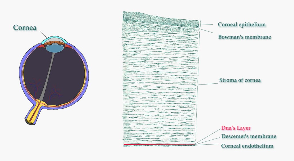
From May 28, 2013 onward, the study of the human eye will forever be changed. A doctor named Harminder S. Dua, Professor of Ophthalmology and Visual Sciences at the University of Nottingham has discovered a new layer of cells that lies just above Descemet’s Layer of the cornea and the corneal stroma. Like so:
“Now hold on there cowboy, what’s the cornea?!”
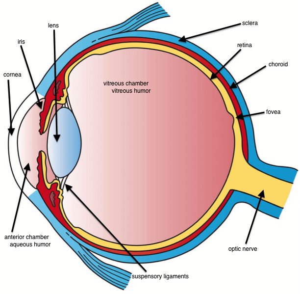
The cornea is the covering for the iris, pupil, and the anterior chamber – basically the spot in front of the eye’s lens. It’s one of the body’s most nerve-filled tissues, and it’s filled with fluid for light transmission. Check this out, it’s an excellent visual description of the cornea, anterior and vitreous chambers — for reference, Dua’s Layer is right between the rear edge of the cornea (closest to the iris) and the middle of the cornea:
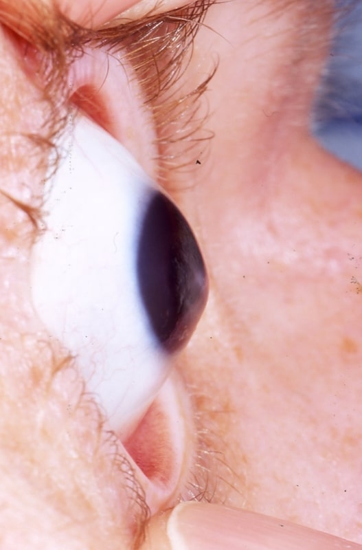
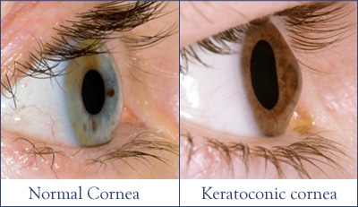
What Dr. Dua has discovered is a layer within the cornea that seems to have something to do with failures in the cornea where misshaping takes place. These kinds of diseases are thought to be caused by water becoming waterlogged within the cornea itself, perhaps caused by a tear in this new Dua’s Layer. They give the person afflicted a cone-shaped cornea that can be corrected with glasses, contacts, or in extreme cases, corneal surgery. I’ve never seen anything quite like this before, so I’m guessing you haven’t either:
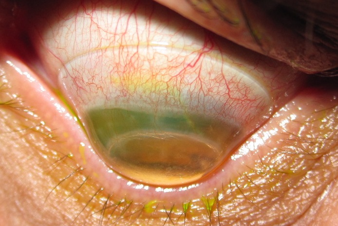
Dua’s Layer is the new tissue discovery that is thought to cause things like this crazy degenerative keratoconus, which looks very annoying and painful to me. Keratoconus causes pretty awful headaches and eye strain for people afflicted, which nobody wants. But, this discovery is being heralded as a potential game changer for corneal diseases and degenerative conditions. From Sci News:
“This is a major discovery that will mean that ophthalmology textbooks will literally need to be re-written. Having identified this new and distinct layer deep in the tissue of the cornea, we can now exploit its presence to make operations much safer and simpler for patients,” said Dr Harminder Dua, Professor of Ophthalmology and Visual Sciences at the University of Nottingham and lead author of a paper published in the journal Ophthalmology.
“From a clinical perspective, there are many diseases that affect the back of the cornea which clinicians across the world are already beginning to relate to the presence, absence or tear in this layer.”
The human cornea is the clear protective lens on the front of the eye through which light enters the eye. Scientists previously believed the cornea to be comprised of five layers, from front to back, the corneal epithelium, Bowman’s layer, the corneal stroma, Descemet’s membrane and the corneal endothelium.
…and from Science Daily:
The scientists proved the existence of the layer by simulating human corneal transplants and grafts on eyes donated for research purposes to eye banks located in Bristol and Manchester.
During this surgery, tiny bubbles of air were injected into the cornea to gently separate the different layers. The scientists then subjected the separated layers to electron microscopy, allowing them to study them at many thousand times their actual size.
Understanding the properties and location of the new Dua’s layer could help surgeons to better identify where in the cornea these bubbles are occurring and take appropriate measures during the operation. If they are able to inject a bubble next to the Dua’s layer, its strength means that it is less prone to tearing, meaning a better outcome for the patient.
The discovery will have an impact on advancing understanding of a number of diseases of the cornea, including acute hydrops, Descematocele and pre-Descemet’s dystrophies.
The scientists now believe that corneal hydrops, a bulging of the cornea caused by fluid build up that occurs in patients with keratoconus (conical deformity of the cornea), is caused by a tear in the Dua layer, through which water from inside the eye rushes in and causes waterlogging.
This is the first time I am ever researching Keratoconus — I have a good friend who has Retinitis Pigmentosa, another degenerative disease of the eye (in that case the retina), but the conical cornea is quite an odd phenomena. Have you ever had or know anyone who has had this disease? I found some information at WebMD on Keratoconus on diagnosis and treatment:
Keratoconus changes vision in two ways:
- As the cornea changes from a ball shape to a cone shape, the smooth surface becomes slightly wavy. This is called irregular astigmatism.
- As the front of the cornea expands, vision becomes more nearsighted. That is, only nearby objects can be seen clearly. Anything too far away will look like a blur.
An eye doctor may notice symptoms during an eye exam. You may also mention symptoms that could be caused by keratoconus. These include:
- Sudden change of vision in just one eye
- Double vision when looking with just one eye
- Objects both near and far looking distorted
- Bright lights looking like they have halos around them
- Lights streaking
- Seeing triple ghost images
To be sure you have keratoconus, your doctor needs to measure the curvature of the. cornea. There are several different ways this can be done.
One instrument, called a keratometer, shines a pattern of light onto the cornea. The shape of the reflection tells the doctor how the eye is curved. There are also computerized instruments that make three-dimensional “maps” of the cornea.
How Is Keratoconus Treated?
Treatment usually starts with new eyeglasses. If eyeglasses don’t provide adequate vision, then contact lenses may be recommended. With mild cases, new eyeglasses can usually make vision clear again. Eventually, though, it will probably be necessary to use contact lenses or seek other treatments to strengthen the cornea and improve vision.A last resort is a cornea transplant. This involves removing the center of the cornea and replacing it with a donor cornea that is stitched into place.
Congratulations to Dr. Harminder Dua and his team at the University of Nottingham for this amazing discovery!
Keep up the excellent game-changing work, good sir!
Check out the abstract at the journal Ophthalmology.
Thanks to Wikipedia on Keratoconus, Dua’s Layer, Traffic Shaper!




i have keratoconus. i’m 24 and was diagnosed several years ago. it’s much worse in my right eye so i can see pretty well through my left. the catch is that my right eye is dominant though so it screws with my depth perception a bit and can be very distracting, particularly at night.
RGP lenses help a lot but are very annoying to wear depending on how lubricated my eyes are.
god help us all.
-wes
Comments are closed.