Hey, you wanna see the inside of my eye? No, really. The inside of my eye.
I’M SERIOUS!
Check it out:
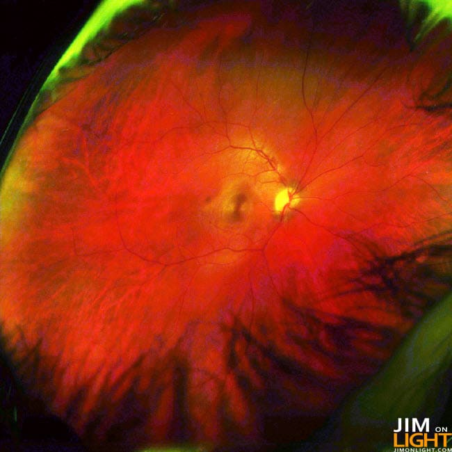
That’s the freaking inside of my right eye – you’re looking at my right retina, optic nerve, macula, and fovea – and a ton of vessels in the background and foreground. Obviously by now you’ve determined that the tree looking things in the bottom of the picture are my eyelashes. Check it out in black and white – around the macula you can see a weird pattern or reflection of some kind – it looks like a lizard eye staring at you!
Oh, is that just me? [awkward]
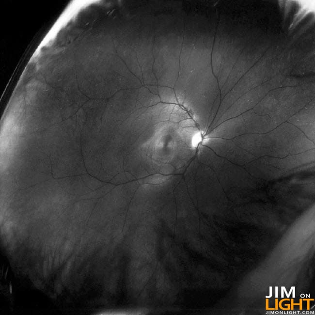
Do you know what the heck I’m talking about? Fovea, macula, retina, etcetera?
If you know all of this already, I am glad to tell you again!
The retina is easy – it’s the large part in the picture. The retina is the back of your eyeball, which contains the light and color receptors (rods and cones, respectively) that the brain uses to tell what’s going on visually. It has blood vessels and stuff like that wound into it so that it can get food and oxygen to the parts of the eye that need it.
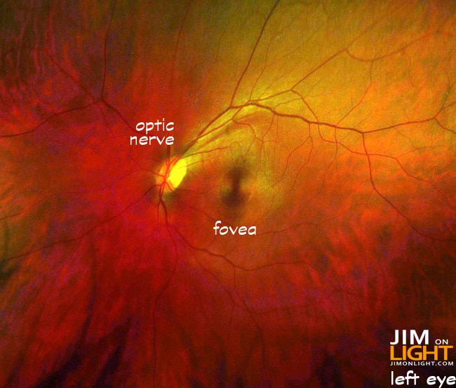
The macula and fovea are an interesting part of your eye. When you hear of “macular degeneration” and people having problems with their visual focus, this is often something to be considered. Check out the left eye – the macula is the spot in the picture below that looks like a violin body, or the mark on the thorax of a Black Widow spider, kind-of. Inside of that is the fovea, which is the central point of focus in our vision:
and even better in black and white:
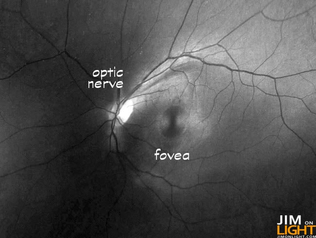
That thing – the fovea – it’s a dip in the retina filled with rod and cone cells, and the center of it is the concentration of human visual acuity, or focus. Around half of the information the optic nerve carries to the brain is from the fovea. The detailed vision spot – when it is damaged, focus goes away. The bright spot is the optic nerve going to the brain, sending messages of everything you see.
The macula is the kind-of yellow-y area surrounding the fovea and containing the fovea – the fovea is essentially the center of the macula.
I always equated the process of sending the images from the eye to the brain like sending a RAW file.
Check out a color shot of my left eye:
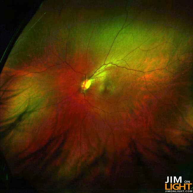
followed by the black and white:
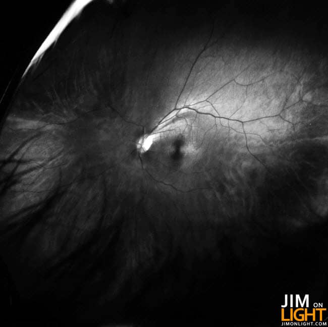
Here’s another term – ischemia. This is a reason to lose weight and be healthy for anyone. An ischemia is a complete lack of blood flow to a portion of the body, and that starved portion dies. Here’s a little game I’ll play – somewhere in one of my eyes I have an ischemia from an old high blood pressure episode. Think you know what it looks like? The first person between now and December 31 who correctly locates the ischemia, I’ll send you a $10 Amazon gift certificate. You have to highlight the ischemia in one of the pictures in this post and email me your guess.
The human body is full of wonder, isn’t it?


